Fig. 3.
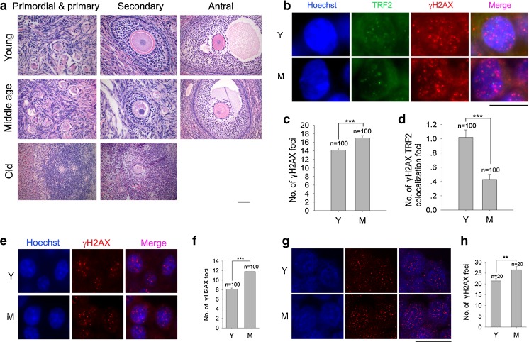
DSBs and telomeres in granulosa cells or cumulus cells of antral follicles in adult monkey ovaries by co-immunostaining of γH2AX and TRF2, or only γH2AX. a Histology showing granulosa cells in the primordial & primary, secondary and mature antral follicles of monkey ovaries. Scale bar = 50 μm; Note, antral follicles are rarely found in old monkey ovaries, so not available for analysis. b Representative morphology of single granulosa cell in antral follicles in the ovaries of young and middle-age monkeys. c Number of γH2AX foci in granulosa cells of antral follicles. d Number of γH2AX and TRF2 co-localization foci in granulosa cells of antral follicles. Scale bar = 10 μm; Error bars show mean + SEM. ***P < 0.001. n = number of cells counted. e-h Immunostaining and confocal imaging analysis of γH2AX foci in the cumulus cells of healthy antral follicles. e Immunostaining images by epi-fluorescence microscopy of γH2AX foci in cumulus cells of follicles in monkey ovaries. f Average number of γH2AX foci in cumulus cells of healthy antral follicles by epi-fluorescence microscopy. g Confocal images by 3-D reconstruction of γH2AX foci in cumulus cells of follicles in monkey ovaries. h Average number of γH2AX foci in cumulus cells of healthy antral follicles by 3-D reconstruction. e Scale bar = 20 μm, (G) Scale bar = 10 μm; Error bars show mean + SEM. **P < 0.01; ***P < 0.001. n = number of cells counted

