
Fig. 1.
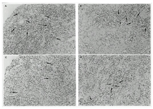
Immunohistochemical staining for p53 in representative synovial tissues from rheumatoid arthritis(A, B) and osteoarthritis(C, D). Positive immunoreactivity appears as brown color(arrows). The amount and pattern of p53 positive cells in rheumatoid arthritis synovial tissues were comparable to those seen in osteoarthritis synovial tissues (Original magnification, X200).