
Fig. 4.
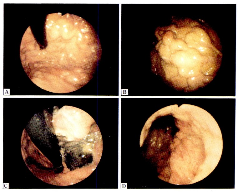
A, B. Endoscopic picture shows a wide spread of several varices over fundus and body. In this case, these varices should be divided into segments in terms of their connection to each other.
C. Endoscopic picture taken 10 days after EVLIS shows necrosis of the gastric varices.
D. Endoscopic picture taken 2 months after EVLIS shows complete eradication of the gastric varices.