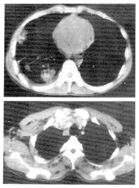
Fig. 4.

Chest CT scan shows an irregular marginated non-enhancing low-density mass with pleural effusion in the left lower lung, and ipsilateral mediastinal lymph nodes enlargement. And round, small, lower-density was detected in SVC, suggestive of thrombosis of SVC.
