Fig. 7.

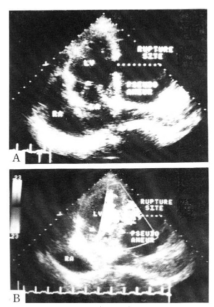
Echocardiogram shows A) a rupture into the pseudoaneurysmal sac from the lateral wall of the left ventricle on apical 4-chamber view, and B) turbulent flow through the rupture site on apical 4-chamber view.

Echocardiogram shows A) a rupture into the pseudoaneurysmal sac from the lateral wall of the left ventricle on apical 4-chamber view, and B) turbulent flow through the rupture site on apical 4-chamber view.