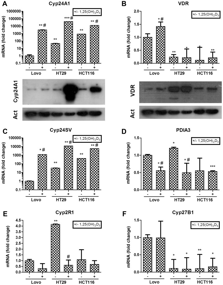
Figure 5.
The effects of 1,25(OH)2D3 on the expression of genes involved in vitamin D3 signaling and metabolism. CRC cell lines were incubated with 0.1 μM 1,25(OH)2D3 for 24 h and the expression of 24-hydroxylase (CYP24A1 gene) (A), vitamin D receptor (VDR gene) (B), splicing variant of 24-hydroxylase (CYP24SV gene) (C), protein disulfide isomerase (PDIA3 gene) (D), 25-hydroxylase (CYP2R1 gene) (E) and 1α-hydroxylase (CYP27B1 gene) (F) was analysed by real-time PCR (qPCR). Data were normalized relative to β-actin mRNA and further normalized against the basal expression in LoVo cells. CYP24A1 (A, lower panel) and vitamin D receptor (B, lower panel) proteins were measured by western blotting, with β-actin used as a control. The data are presented are mean ± SEM. Significance level relative to the LoVo cell basal expression: *P<0.05, **P<0.01, ***P<0.001. Significance level relative to the untreated control for each cell line is indicated as #P<0.05.

