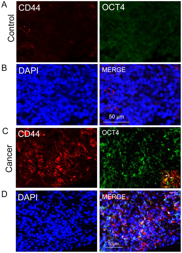
Figure 1.
CD44-positive cancer cells in NPC tissues and nasopharyngitis epithelial cells. (A) CD44 (red) was expressed in only a few nasopharyngitis epithelial cells, and OCT4 was rarely detected in these cells. (B) The merged image shows little co-expression of CD44 and OCT4 (green) in nasopharyngitis epithelial cells. (C) CD44 and OCT4 were expressed in select regions of NPC tissue sections, mainly in epithelial areas instead of lymph regions. Double-immunolabeling showed that some regions contained co-expression of CD44 and OCT4. (D) Cell nuclei were counterstained with DAPI (blue), and the right panel shows the merged color. The magnification is 50 μm.

