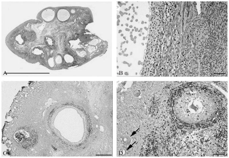
Figure 2.
Ovarian histology in a biopsy proven case of autoimmune oophoritis. Hematoxylin and eosin staining shows multiple antral follicles in patient 1 (A), bar = 1 cm. Higher magnification shows lymphocytic infiltration of the theca of an antral follicle and luteinized granulosa cells (B), bar = 50 μm. Immunoperoxidase staining for CD3 highlights infiltration of lymphocytes into the theca in this patient (C), bar = 500 μm. Immunoperoxidase staining for CD3 demonstrates infiltration of lymphocytes into the theca of a preantral follicle in patient 4 and presence of earlier stage follicles free of lymphocytic infiltration (arrows) (D), bar = 100 μm. [From Bakalov VK et al.].49

