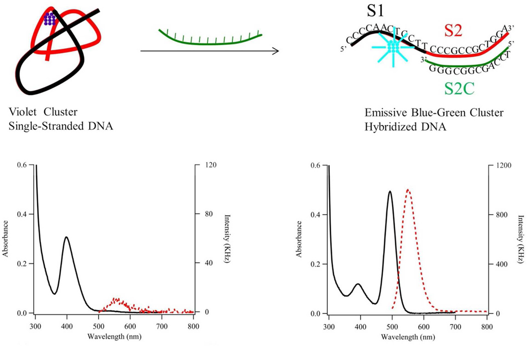
Figure 1. Reaction Scheme.
Our proposed reaction scheme describes how hybridization changes both DNA structure and silver cluster spectra. (Left) The composite strand has the 5’ cluster domain (S1 = CCCCAACTCCTT (black segment)) and one of five recognition sites (S212a = CCCGCCGCTGGA (red segment)). This single-stranded oligonucleotide hosts an ~11 silver atom with λmax = 400 nm (solid black line – left axis) and low emission (dashed red line – right axis). The cluster condenses its host strand. (Right) This cluster-laden strand hybridizes with its complementary strand (S2C12a = TCCAGCGGCGGG (green segment)). Cluster size is conserved but λmax shifts to 490 nm (solid black line – left axis) and strong green emission develops (dashed red line – right axis with 10X scale difference).

