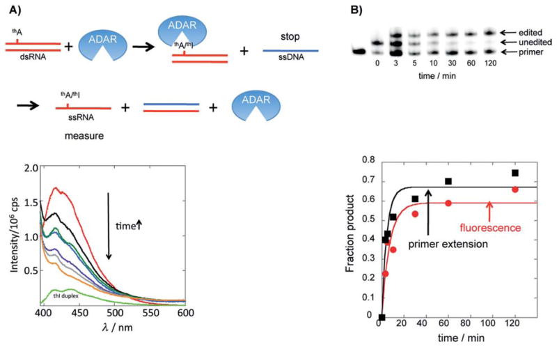
Figure 5.
Fluorescence-based assay for an ADAR reaction. A) Top: The procedure used to stop the reaction such that thA/thI fluorescence can be measured in ssRNA form. Bottom: Plot of change in fluorescence as a function of time for the reaction of 1 mm thA-modified GluR B R/G site substrate and 1 mm hADAR2-RD. Red: 0 min, black: 3 min, blue: 5 min, dark green, 10 min, purple: 30 min, gray: 60 min, orange: 120 min, light green: thI-containing duplex. B) Top: Image of gel used to resolve products for primer extension editing assay for reaction evaluated by changes in fluorescence in (A). Bottom: Fraction product vs time for primer extension assay (black squares) and fluorescence assay (red circles) (kobs=0.14 0.01 min¢1 (fluorescence); kobs=0.15 0.07 min¢1 (primer extension).

