
Figure 2.
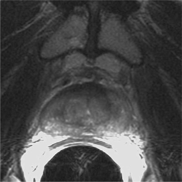
63 year-old man with biopsy-proven recurrence of prostate cancer in the right mid-gland and base 4 years and 2 months after treatment. Axial T2-weighted image (TE/TR 5000/96) shows diffuse low signal intensity in the peripheral zone and central gland. Both readers interpreted it as a negative case.