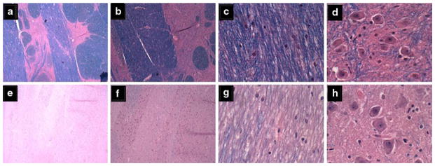
Fig. 4.

Central pontine myelinolysis. a–d Control basis pontis showing a–c intact myelin, a, b clear delineations between gray and white matter structures, and d abundant pontine neurons. e–h Alcoholic basis pontis (same as Fig. 7a) showing e–g almost complete loss of myelin, e, f virtually no delineation between gray and white mater structures, and b reduced abundance of glial cells, yet h preservation of neurons in pontine neurons (Luxol Fast Blue, Hematoxylin and Eosin stain)
