
Fig. 7.
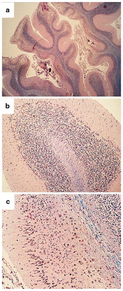
Alcoholic cerebellar degeneration. a Pronounced atrophy of cerebellar folia in the anterior vermis marked by widening of fissures and thinning of cortical and white matter structures. b Higher magnification of the cortex showing attenuation of cells in the inner granule cell (igc) layer, and proliferation of Bergmann’s glia (band-like; arrowheads) with loss of Purkinje cells. c A higher magnification image depicting severe degeneration of the cortex with subtotal depletion of neurons in the granule and Purkinje cell layers, and fiber loss in white matter cores (blue, center right). At the far right, the granule cell population is reduced but better preserved compared with the left side of the same folium