
Fig. 10.
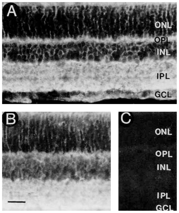
A: GAT-3 immunoreactivity is distributed to all regions of the retina. The immunostaining pattern indicates that it is predominantly expressed by Müller cells. B: No change in GAT-3 immunostaining in a section incubated with GAT-3 antibody preadsorbed with 10−5 M GAT-1588–599. C: Lack of GAT-3 immunostaining in a control section incubated with the GAT-3 antibody preadsorbed with 10−5 M GAT-3607–627. Scale bar = 25 μm.