
Fig. 11.
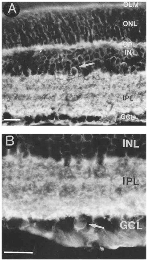
A: GAT-3 is predominantly expressed by Müller cells, but it is also expressed by some amacrine and displaced amacrine cells. A GAT-3–immunoreactive amacrine cell (arrow) in the proximal INL. B: A GAT-3–immunostained displaced amacrine cell (arrow) in the GCL. Abbreviations as in Figure 1. Scale bar = 25 μm.