
Fig. 3.
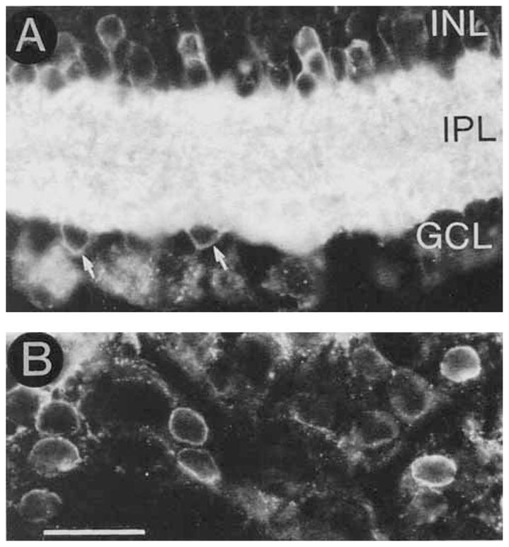
GAT-1 immunoreactivity in the INL and GCL. A: Transverse section of the retina illustrating small GAT-1–immunoreactive cell bodies in the proximal INL and GCL. Arrows indicate GAT-1–containing displaced amacrine cells. B: Horizontal section of the retina through the GCL. GAT-1 immunostaining is predominantly localized to the plasma membrane of many small cell bodies. These small cells are displaced amacrine cells. Some diffuse immunostaining of Müller cell processes between these cells can he seen in this figure. Scale bar = 25 μm.