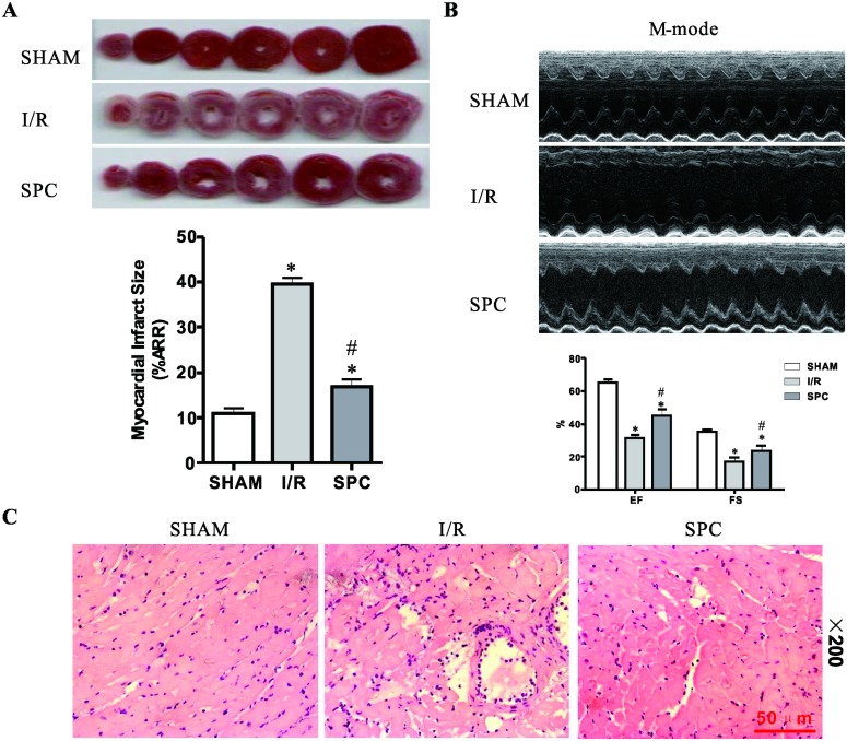
Fig 1. SPC decreases cardiac infarct size, increases LV contractile function and attenuates histopathologic organ damage following I/R.
(A) Rats were sacrificed at the end of reperfusion and the hearts removed and stained with TTC for the measurement of myocardial infarct area. The infarct size was expressed as a percentage of area at risk. n = 6 /group. (B) Echocardiography was performed at the end of reperfusion and representative M-mode echocardiograms were recorded in all groups. Rats without LAD occlusion served as basal controls (SHAM group). n = 10 /group. (C) LV tissues were retrieved at the end of reperfusion, paraffin section was prepared and subjected to the H&E staining. Representative H&E staining images are shown (magnification, 200×). Scale bar: 50μm. n = 3 /group. The columns and errors bars represent means ± SD. * P < 0.05 vs. SHAM group; # P < 0.05 vs. I/R group.

