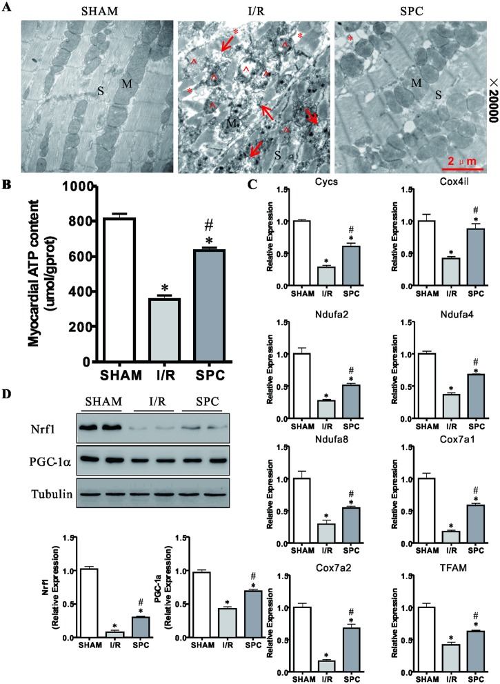
Fig 2. SPC ameliorates mitochondrial disorder and dysfunction after I/R.
(A) LV tissues were harvested for examination of myocardial ultrastructure by transmission electron microscopy (TEM). Typical TEM images obtained at a magnification (20000×) of cardiac ultrastructure in all groups. Note that myofilaments were absent (*) and damaged mitochondria (^) nearby the autophagosomes (→). Scale bar: 2μm. n = 3 /group. M: mitochondria; S: sarcomeres. (B) SPC prevents depletion of ATP stores in I/R hearts and the ATP content in all groups are shown. n = 6 /group. (C) After 2 h of reperfusion, LV tissues were obtained and analyzed by real-time PCR for the expression levels of Cycs, Cox4il, Ndufa2, Ndufa4, Ndufa8, Cox7a1, Cox7a2 and TFAM. n = 6 /group. (D) LV were collected and prepared for immunoblots. Representative immunoblots and semiquantitative analysis of Nrf1 and PGC-1 in each group of rats after reperfusion. The blots for Tubulin were served as loading controls. n = 4 /group. All data are presented as means ± SD. * P < 0.05 vs. SHAM group; # P < 0.05 vs. I/R group.

