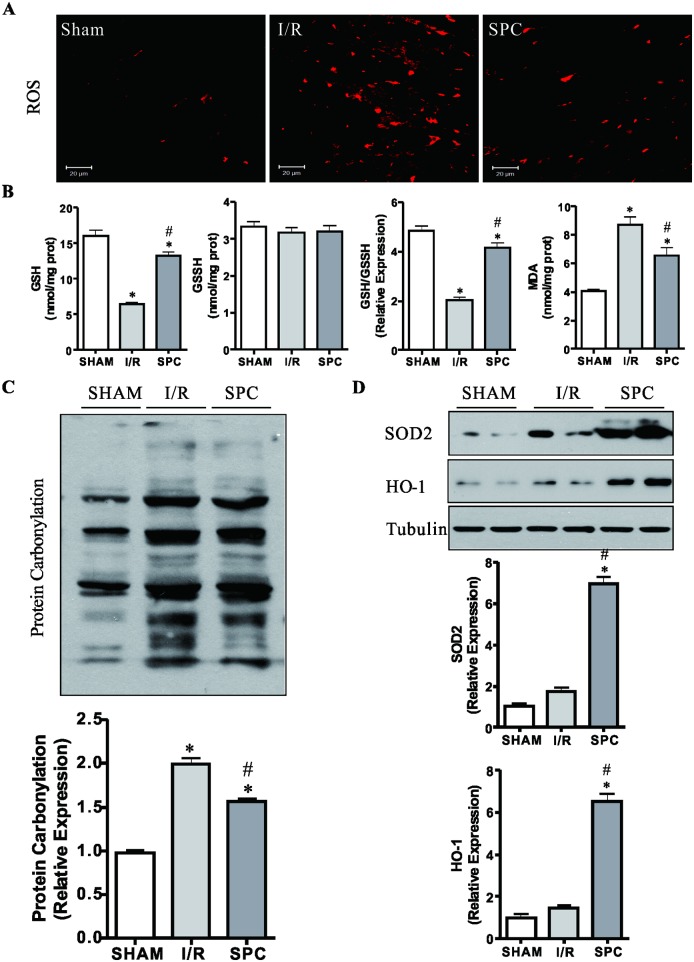
Fig 3. SPC inhibits oxidative stress brought about by the I/R injury.
(A) The cardiac levels of ROS in each group are shown by DHE staining. The image was obtained by a confocal microscope. SPC-treatment significantly lowered the increased DHE fluorescent intensity induced by the I/R injury. Scale bar: 20μm. n = 3 /group. (B) The cardiac GSH, GSSG and MDA levels were measured using enzymatic kits, and the GSH/GSSG ratios derived from the GSSG and GSH contents. n = 6 /group. (C) Protein carbonyl content was examined as carbonyl-containing 2,4-DNPH adducts by immunoblotting. Protein carbonylation of SPC group was likewise lower than the I/R group. n = 4 /group. (D) The immunoblot analysis SOD2 and HO-1 expression at the end of reperfusion. The blots for Tubulin were served as loading controls. n = 4 /group. The columns and errors bars represent means ± SD. * P < 0.05 vs. SHAM group; # P < 0.05 vs. I/R group.

