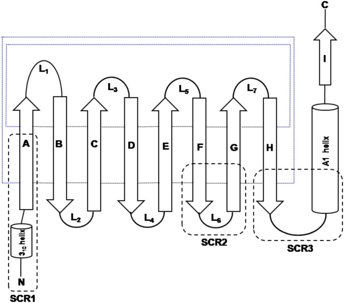
Fig 1. Topology diagram of the lipocalin fold.
The strands [A-I] of the β-sheet and the helices are represented as arrows and cylinders respectively. The loops connecting the strands are marked as L1-L7. The hydrogen bond connections between the strands are shown in small dotted lines. The three structurally conserved regions (SCR) are marked as SCR1, SCR2 and SCR3 respectively. Blue lines indicates that β-strands A and H are in-register and thus forming a β-barrel structure. Black lines indicates that adjacently shown strands are in-register.

