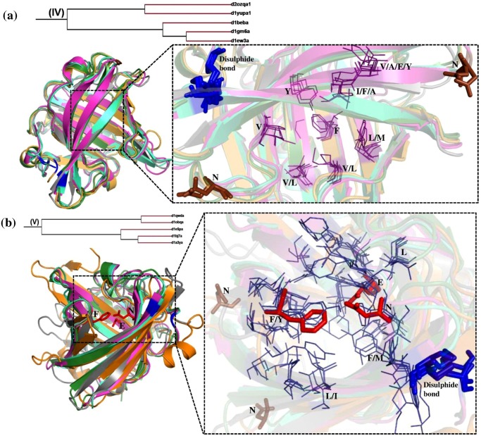
Fig 7. Structural superimposition of lipocalin domains in cluster IV (a) and cluster V (b) corresponding to Retinol protein binding protein-like family.
(a) Structural superimposition of Bovine β-lacto globulin (d1beba_) (green), salivary lipocalin (d1gm6a_) (cyan), Major horse allergen (d1ew3a_) (pink), Major urinary protein (d2ozqa1) (grey) and Reindeer β-lacto globulin (d1yupa1) (orange). (b) Structural superimposition of Pheromone binding protein (d1e5pa_) (pink), Lipocalin allergen (d1bj7a_) (cyan), Odorant binding protein (d1a3ya_) (green), Lipoprotein Blc (d1qwda_) (grey) and α-crustacyanin (d1obqa_) (orange). The ligand binding region is represented as dotted line box. The residues in the binding regions are shown as purple lines in the closer view. The ligand interacting residues are represented as red sticks. The disulphide bond is represented as blue sticks. Asn that is involved in glycosylation are shown as brown sticks.

