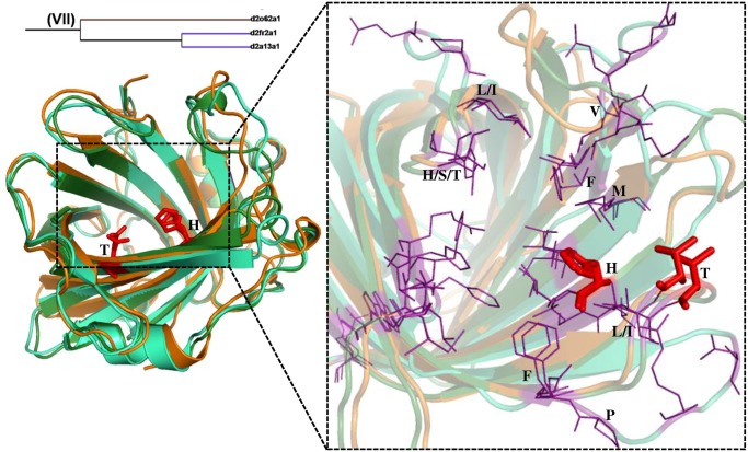
Fig 9. Structural superimposition of lipocalin domains in cluster VII corresponding to RV2717c-like and all 1756-like families.
The ligand binding region is represented as dotted line box. The residues in the binding regions are shown as purple lines in the closer view. The ligand interacting residues are represented as red sticks. Shown are the superimposition of Uncharacterized protein DUF 3598 (Nostoc punctiforme) (d2o62a1) (cyan), Rv2717c DUF 1794 (M. Tuberculosis) (d2fr2a1) (orange) and Nitrophorin-like heme binding protein (Arabidopsis Thaliana) (d2a13a1) (green).

