
Figure 2.
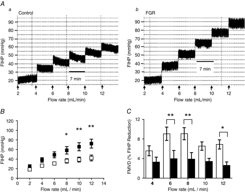
Vascular resistance in placentas from normal and fetal growth restricted (FGR) pregnancies
A, ex vivo dually perfused placental lobules were subjected to staged (7 min) incremental increases in fetal-side perfusate flow and fetal-side inflow hydrostatic pressure (FIHP) measured as an indication of vascular resistance. Traces are representative of FIHP in normal (Aa) and FGR (Ab) placentas. B, mean resistance values in placental lobules from normal (n = 10; open squares) and FGR (n = 10; filled squares) pregnancies were obtained for all flow rates during steady-state, 6–7 min following alterations in flow. Two-way ANOVA: P < 0.0001; Bonferonni test: *P < 0.05, **P < 0.01. C, flow-mediated vasodilatation (FMVD) was measured as the percentage decrease in FIHP, from peak FIHP to the new steady state following an elevation in flow, in normal (open bar) and fetal growth restricted (filled bar) placentas. Data show means ± SEM; 2-way ANOVA: P < 0.001; Bonferonni test: *P < 0.05, **P < 0.01.