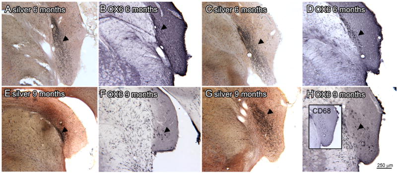
Figure 11.

Silver staining and OX6 expression at 6 (A-D) and 9 (E-H) month survival times. A. Silver staining in VCP (arrowhead; bregma -11.195). B. An adjacent section showing OX6 expression within but smaller than the region of silver staining. C. Silver stained section through VCP and CN, the arrowhead indicates the region of darkest staining (bregma -11.096). D. Slightly more caudal section showing minimal OX6 immunolabel (arrowhead; bregma -11.195) E. Silver staining in DC (arrowhead; bregma -11.756). F. The arrowhead indicates a few OX6-ir elements on an adjacent section. G. Silver staining in the VCP (bregma -11.063). The inset shows CD68-r elements on an adjacent section. H. The arrowhead indicates an OX6-ir element on an adjacent section.
