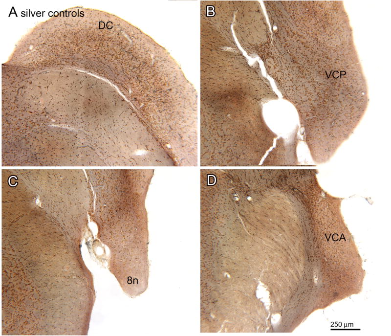
Figure 2.

Absence of silver staining in the CN of control animals. A. Section through the DC (bregma -11.558). B. Section through the VCP (bregma -11.195). C. Section through the 8n and VCP (bregma -10.568). D. Section through the VCA (bregma -9.842). Scale bar also for A, B, C.
