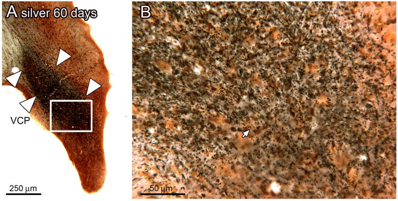
Figure 3.

A. Dense silver staining in the VCP of an experimental animal, 60 day survival time. The white arrowheads mark the dense band of silver staining in the VCP (bregma -10.997). The rectangle shows the location of the higher magnification image in B. B. The staining is made of of black puncta, some of which are arranged in a line (arrow).
