
Figure 6.
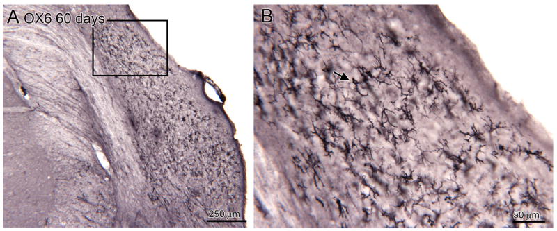
A. Distribution of activated microglia in the CN of an experimental animal, 60 day survival (bregma -10.898). The rectangle shows the location of the image in B. B. The labeled elements have a variety of shapes, some with thick processes (example at arrow), others with thin processes.