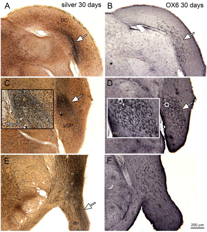
Figure 7.

Silver staining and OX6 expression, bilateral noise exposure, 30 day survival. A. Band of silver staining (arrow) on a section through the DC (bregma -11.591). B. Adjacent staining showing a limited region of OX6 expression (white arrow). C. Silver staining (white arrow) in VCP (bregma -11.723). The asterisk is an alignment point for the inset. The inset shows a higher magnification image of the silver staining, showing dense black puncta. D. Adjacent section showing OX6 expression in the VCP. The asterisk is an alignment point for the inset that shows the appearance of the activated microglia. E. Silver staining (arrow) in the eighth nerve (bregma -11.096). F. Scattered OX6-ir profiles on an adjacent section.
