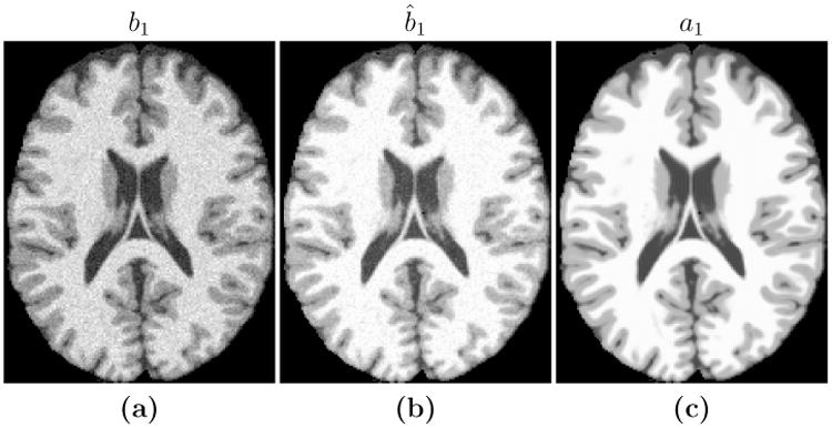
Figure 3.

Shown are (a) an SPGR image (TR = 18ms, TE = 10ms, α= 45° with 3% additive noise) which we use as our subject image b1, the (b) reconstruction, b̂1, of the subject image with the same pulse sequence as used to image (c) the atlas target image a1 (TR = 18ms, α= 30°, TE = 10ms with 0% noise).
