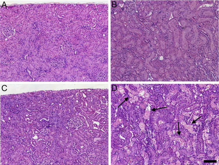
Fig. 9.

Histological analyses of kidneys from pregnant Wistar dams injected intravenously with 0.5 μmol HgCl2 kg−1 2 mL or 2.5 μmol HgCl2 kg−1 2 mL. In kidneys of rats injected with 0.5 μmol HgCl2 kg−1 2 mL, no histological alterations or pathological changes were observed in the cortex (A) or outer stripe of the outer medulla (B). In kidneys of rats injected with 2.5 μmol HgCl2kg−1 2mL, the cortex (C) appeared normal; however, significant areas of necrosis (arrows) were evident in the outer stripe of the outer medulla (D). Bar= 100 μm.
