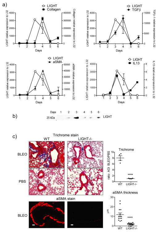
Figure 1. LIGHT is induced by bleomycin and LIGHT-deficient mice exhibit decreased lung fibrosis.
(a–b) Wild type (WT) mice were sensitized with bleomycin intratracheally (i.t.). (a) Kinetics of expression of mRNA for LIGHT, alpha smooth muscle actin (aSMA), TGF-β, and IL-13, assessed by qPCR analysis of lung samples. mRNA expression calculated relative to L32. Values are mean ± SEM of 2 mice per time point. Selected time points were repeated in 3 additional experiments. (b) Soluble LIGHT was assessed in the bronchoalveolar lavage by Western Blot. Data are representative of 3 experiments. (c) WT and LIGHT−/− mice were sensitized with bleomycin or PBS intratracheally. Mice were sacrificed on day 7. Fibrotic phenotypes were assessed by analyzing collagen deposition (trichrome stain) and peribronchial aSMA accumulation in lung sections. Representative sections are shown (left). Quantitation was performed as in materials and methods (right). Trichrome data show mean ratio of area of interest (AOI) values over PBS of 4 individual mice per condition. aSMA data show values of 14 random bronchi scored from 4 mice per condition. Data are representative of 6 experiments.

