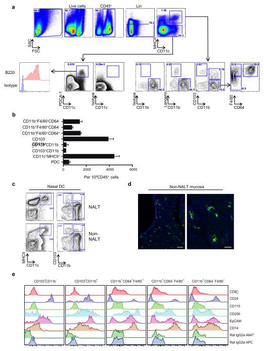
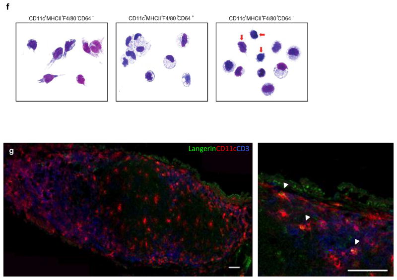
Figure 2. Phenotypic characterization of nasal DCs.
(a, b, c) Single cell suspension of nasal cells was obtained by mechanical disruption and collagenase-digestion of nasal tissue. Multiparameter flow cytometry was used to identify DC subsets within the murine nose. (a) Defines the gating strategy used to delineate myeloid DC (mDC) subsets based on the expression of CD103, CD11b, and CD64 on live, CD45+lineage−CD11c+MHCII+ cells. For identification of plasmacytoid DCs (pDCs), MHCII− cells were defined on the basis of CD11c and PDCA-1 staining. PDCA-1hiCD11c+B220+ cells were deemed putative pDCs. (b) Absolute number of all DC subsets including pDCs. Error bars = standard deviation (SD). (c) Shows identification of NALT and non-NALT DCs separately and (d) Shows eYFP+ cells in non-NALT mucosa. Bars, 50μm (left) and 25μm (right). (e) Shows detailed phenotypic characterization of the nasal APC subsets using a panel of monoclonal antibodies. (f) Wright-Geimsa stains were used to define the cellular morphology of putative nasal DCs and nasal Mϕ. Bars, 10μm. (g) CD11c+langerin+DCs were analyzed by immunofluorescence using confocal microscope where langerin+ cells are depicted in green, CD11c+ cells in red and CD3+ cells in blue. White arrowheads indicate the cells where co-localization of langerin with CD11c was observed. Bars, 50μm.


