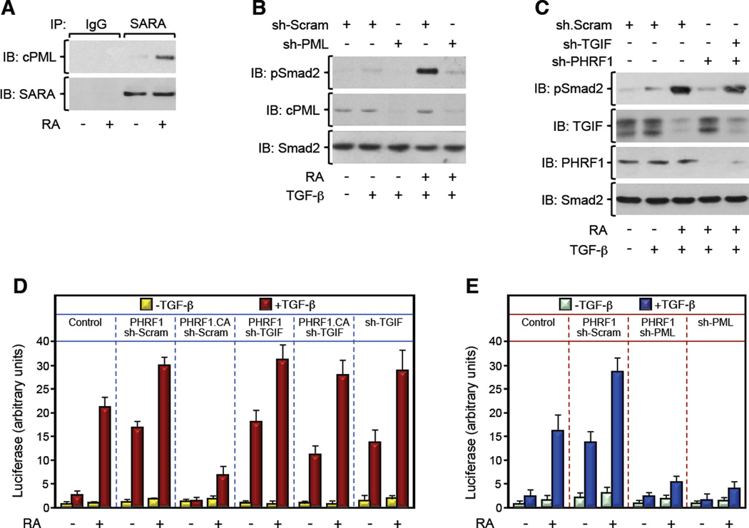
Figure 3. PML-RARα Suppresses TGF-β Signaling by Disrupting PHRF1-Driven TGIF Degradation. See also Figure S3.
(A) NB4 blasts were treated with RA for 24 hr, and the association of endogenous cPML and SARA was analyzed by coimmunoprecipitation.
(B, C) NB4 blasts expressing the indicated combinations of sh-RNAs were cultured with RA for 24 hr. Then, cells were treated with TGF-β for 30 min and phosphorylation of Smad2 was analyzed by immunoblotting using anti-phospho-Smad2 (pSmad2) antibody.
(D, E) NB4 blasts were transfected with ARE3-Lux together with FAST1 and the indicated combination of expression vectors and treated with RA for 24 hr before being treated with TGF-β for the last 16 hr. Luciferase activity was measured and data are expressed as mean ± SD (n = 3).

