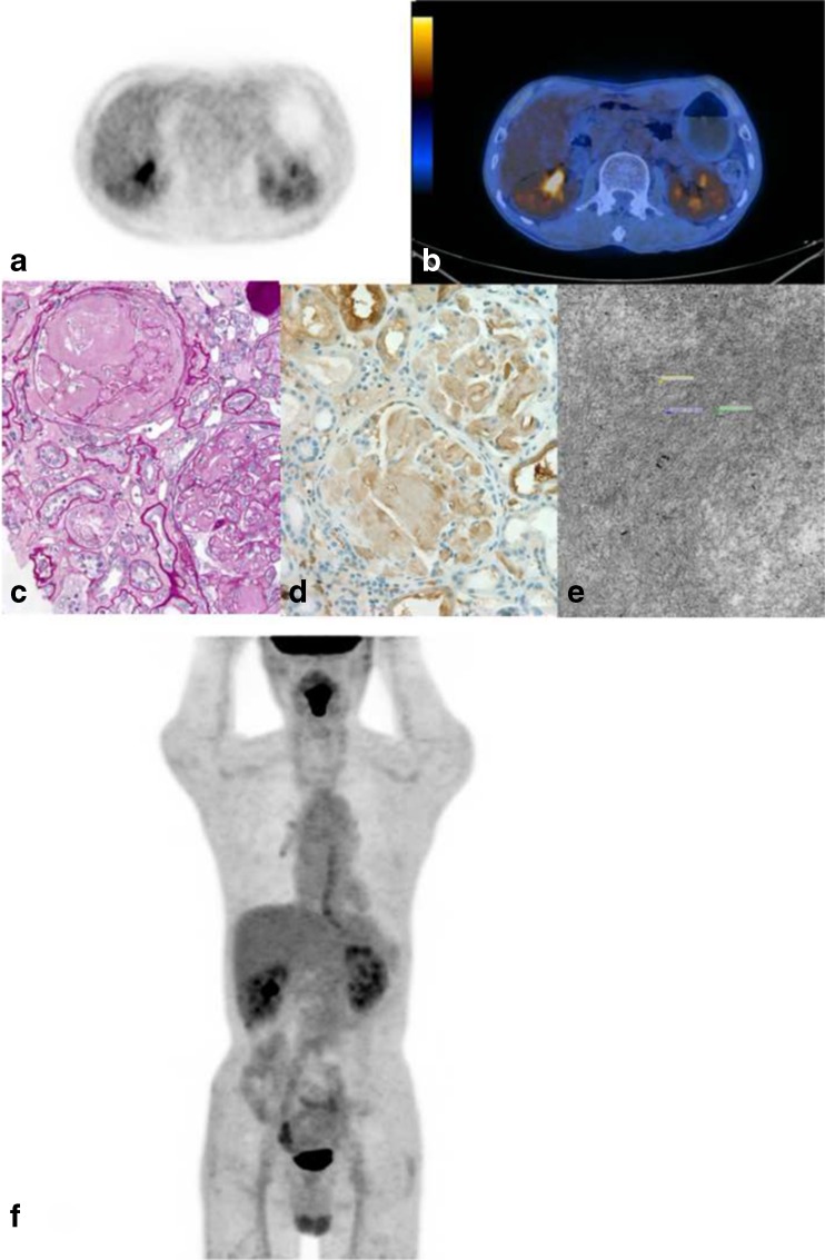
Fig. 2.
Abnormal diffuse renal parenchymal 18F-FDG uptake (SUVmax = 3.5) on transverse PET (a), fused PET/CT images (b) and MIP (f) in a 61-year-old male patient with biopsy-proven bilateral renal amyloidosis. Pathological specimen obtained by renal biopsy revealed amyloid deposition in periodic acid Schiff staining (c), immunohistochemical staining for anti-lambda antibody (d) and electron microscopy showing interlacing bundles of parallel arrays of fibrils with a diameter of 11–13 nm (e)

