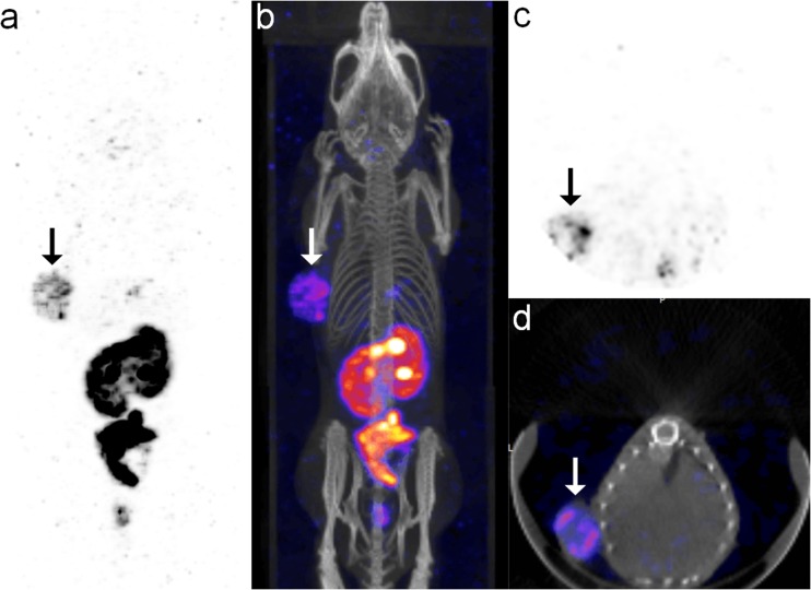
Fig. 5.
Micro-SPECT/CT images of KB tumor-bearing mouse obtained at 22 h after intraperitoneal administration of 99mTc-ECG-EDA-folate. a, b MIP and fusion images show clear visualization of an FR-positive KB tumor in the left axilla (arrows). c, d Transaxial and fusion images of the KB tumor show rim-shaped tumor uptake with central photon defect

