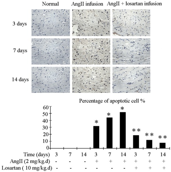
Figure 1.

Rat atrial tissue apoptosis detection with the TUNEL procedure. Upper panel show representative photograph of TUNEL. Nucleiof normal cell were stained blue, while nuclei of apoptotic cell were stained brownish black. Bottom panel shows quantification by percentage. Ang II infusion group demonstrated higher level of apoptosis compared to the control group. This increase in apoptosis was attenuated in Ang II + losartan group. n = 3 animals per group; data are mean ± SD. *P < 0.05 vs control group, **P < 0.05 vs Ang II infusion group.
