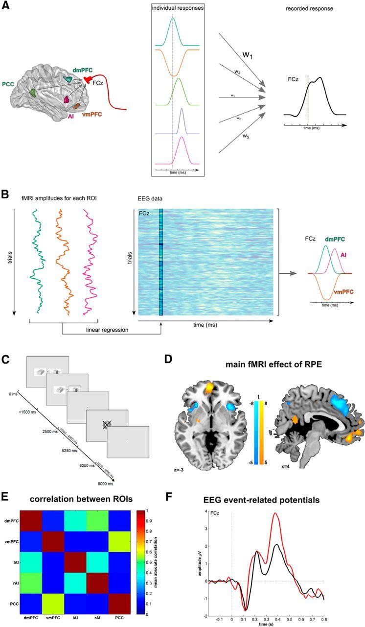
Figure 1.

Contributions of multiple brain areas to the EEG signal and their parcellation using fMRI-informed EEG. A, fMRI studies of reward processing show a multitude of areas responsive to reward information, such as reward prediction errors (left). Hypothetically, each area has a specific time course and amplitude in response to a stimulus (middle). Electrical fields elicited by these individual activation patterns sum spatially and a mixture of these signals defines event-related potentials measured at scalp electrodes, such as frontocentral electrode FCz (right). The contribution of each area is governed by the strength of its activation and the spatial proximity to the electrode, such that the recorded signal is a weighted sum (as indicated by hypothetical weights, w1–w5) that overweighs sources close to the recording electrodes (here: dmPFC). B, Trial-by-trial fMRI-informed EEG analysis disentangles unique contributions of brain areas to the EEG signal. Using the hemodynamic response, we estimate fMRI activation of each brain region on every trial (left). Within a multiple-regression analysis, we then determine the peristimulus time when trial-by-trial fMRI activations best predict the EEG signal (middle). This enables us to recover the unique and individual time courses of these brain regions (right). However, a caveat is that the approach remains insensitive to temporal contributions that perfectly covary in the fMRI signal across trials. C, Participants engage in a probabilistic reversal-learning task where they learn which of two stimuli had the higher reward probability. One of the stimuli was assigned as the correct stimulus with a reward probability of 80%. The other stimulus had a reward probability of 20%. The reward was depicted by a framed 50 Swiss Centimes' coin, whereas a punishment was illustrated by a crossed coin. After 6–10 correct responses, the reward probabilities reversed. The participants were informed about potential reversals but were not instructed on the timing of these reversals. D, fMRI activations for RPEs at feedback. Increasing RPEs were linked to engagement of vmPFC and PCC, whereas decreasing RPEs engendered activation in dmPFC and bilateral AI. E, Average absolute correlations between ROIs activated in the RPE-fMRI-contrast. F, Event-related potentials elicited over electrode FCz, separated for rewards (black) and punishments (red). dmPFC, Dorsomedial prefrontal cortex; lAI, left anterior insula; PCC, posterior cingulate cortex; rAI, right anterior insula; vmPFC, ventromedial prefrontal cortex.
