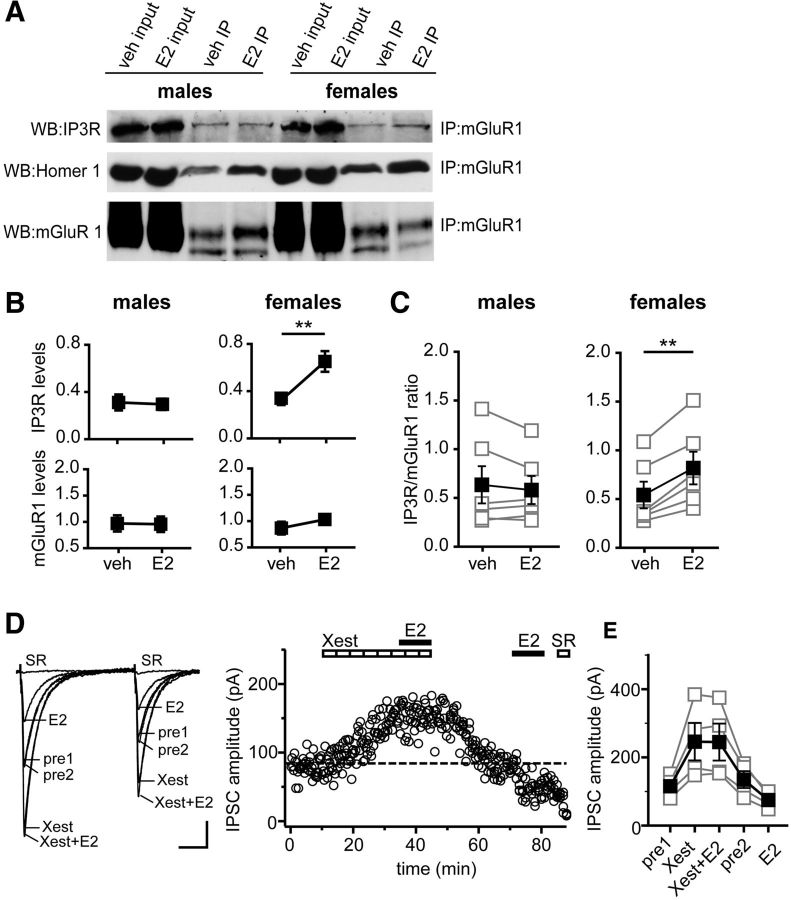
Figure 6.
E2 acutely promotes mGluR1–IP3R interaction in females but not males. A, Hippocampal slices from male and female rats were treated either with E2 (100 nm) or vehicle (veh) for 10 min. Membrane fractions were prepared from these slices and subjected to IP using anti-mGluR1a. Representative Western blots (WB) of input and IP samples from males and females on the same gel are shown. The blots were probed for IP3R, Homer-1b/c and mGluR1. B, Mean ± SEM of IP3R and mGluR1 levels in males and females (n = 6 independent experiments). E2 increased the levels of IP3R associated with mGluR1a in females, but not in males, without affecting mGluR1a levels in either sex; **p < 0.01, Student's t test. C, IP3R/mGluR1 ratio for the same six experiments as in B (see Materials and Methods). Connected open symbols are individual experiments (n = 6); filled symbols are mean ± SEM for all experiments. E2 increased the IP3R/mGluR1 ratio in females with no effect in males; **p < 0.01, paired t test. D, Individual traces and time course of IPSC suppression in a representative experiment in which E2 (100 nm) was applied first in the presence of Xest (IP3R inhibitor, 2 μm) and then again after Xest washout to confirm E2 responsiveness of IPSCs. Dotted line shows average IPSC amplitude during 2 min before the second E2 application. Xest blocked E2-induced IPSC suppression. Each point in the time course is an individual sweep, and SR 95531 (SR; 2 μm) applied at the end of the experiment blocked IPSCs. E, Group IPSC amplitude data for all experiments with E2-responsive IPSCs (n = 4). Connected open symbols are individual cells; filled symbols are mean ± SEM for all cells. Calibration: D, 25 pA, 25 ms.

