Figure 5.
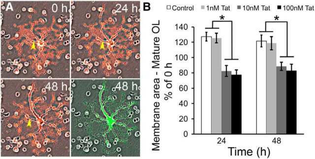
Tat reduces myelin-like membranes of mature OLs. A, Sample images showing changes in mature OL membrane area over 48 h. Cells were labeled with 2 μg/ml CM-DiI 1 d before Tat treatment. Phase contrast and fluorescent images of the same cell were taken at 0, 24, and 48 h. Phase and fluorescent images were merged to better visualize the myelin-like membrane structures. At the end of the experimental period, cells were labeled with calcein-AM to verify viability (green fluorescence). The yellow arrowhead indicates an area with an obvious reduction in the area of the cellular processes (loss of red fluorescence) over time. B, Quantification of the change in membrane area in vehicle and Tat-treated OLs. At both 24 and 48 h, vehicle-treated OLs showed ∼25% growth of membrane area. Tat (1 nM) did not have any significant effect on the membrane area when compared with controls. However, 10 or 100 nm Tat-treated OLs at 24 and 48 h exhibited ∼20% reductions in membrane area compared with 0 h (*p < 0.05, 2-way ANOVA followed by post hoc Bonferroni's test; n = 4 individual experiments; ≥25 cells were counted for each n).

