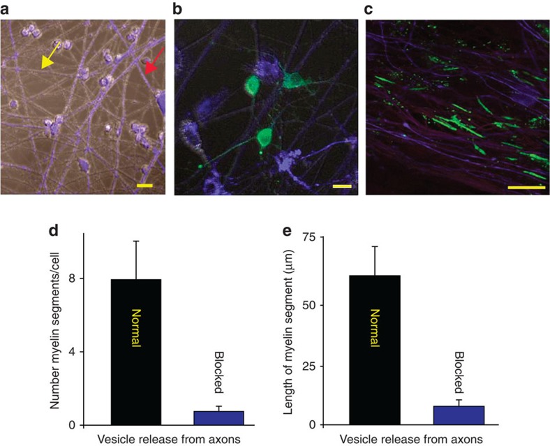
Figure 1. Electrically active axons releasing synaptic vesicles are preferentially myelinated.
(a) DRG neurons treated with BoNT/A and stained with cell tracker (blue, see red arrow) co-cultured with normal (untreated) neurons (grey, see yellow arrow). (b) OPCs (green, GCaMP3) were added to the cultures to determine whether exocytosis of neurotransmitter from axons influenced myelination. (c) Axons were stimulated for 9 s at 10 Hz every 5 min for 10 h and cultured for 3 weeks. Myelin (green, myelin basic protein, MBP) analysed 3 weeks after co-culture formed preferentially on axons releasing synaptic vesicles (purple, neurofilament), and (d) number of myelin segments/cell were more in normal axons (P<0.001, n=7 cells from four dishes) (e) myelin segments were also longer in normal axons (P<0.001, n=9 cells from four dishes). Scale bar, 10 μm (a,b); 20 μm (c).

