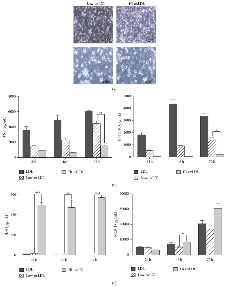
Figure 5.
Morphological changes and cytokine production in primary monocytes. (a) Human blood monocytes were isolated from PBMCs and cultured with both M-CSF (50 ng/mL) and differentially oxidized LDL (50 μg/mL each) for 7 days. Morphological changes were observed using bright-field microscope (×200, 400). (b) Primary monocytes were pretreated with both M-CSF (50 ng/mL) and differentially oxidized LDL (50 μg/mL) for 24, 48, or 72 h and treated with LPS (20 ng/mL) for 18 h. Culture supernatants were harvested and the levels of TNF-α, IL-12p40, IL-6, and MCP-1 were measured by ELISA. Data represent means ± S.E. and one-way ANOVA analysis was used to determine significance (∗ p < 0.05, ∗∗ p < 0.01, and ∗∗∗ p < 0.001).

