Figure 2.
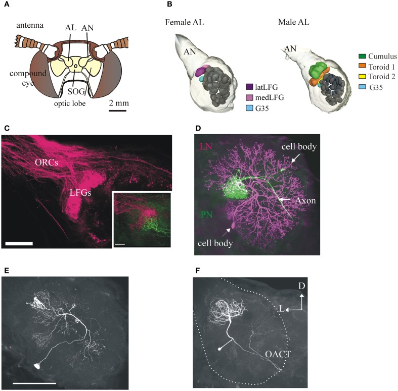
Anatomy of the primary olfactory center of the moth M. sexta and its neuronal components. (A) Schematic frontal view of the moth's head. Each antennal lobe (AL) receives input from antennal olfactory sensory neurons (OSNs) through the antennal nerve (AN) (one glomerulus receives input from the labial palps, not shown). SOG, suboesophageal ganglion. (B) 3-D reconstructions of the M. sexta ALs from a female (left) and a male (right), showing the sexually dimorphic glomeruli (LFGs in females, the Cumulus and the two Toroids in males) and a sexually isomorphic glomerulus (G35, in light blue) of known odor input. Scale bar: 50 μm. (C) Confocal image showing OSN afferent input to the LFGs. Inset: mass-labeled OSNs with a single-labeled LFG projection neuron (in green). Scale bar: 100 μm. Figure with permission from Dr. J. Hildebrand. (D) A female AL showing the two main types of neurons, a uniglomerular projection neuron (PN, in magenta) and a local interneuron (LN, in green). PNs have an axon that projects from the AL to higher brain centers in the protocerebrum; LNs are intrinsic to the AL and connect many glomeruli. Scale bar: 100 μm. (E) LNs are heterogeneous. A LN with arborizations restricted to relatively low number of glomeruli (compare with the LN in D). Scale bar: 100 μm. (F) The ALs also contain a sizable number of multiglomerular PNs (Homberg et al., 1988), whose functions have not been systematically studied. This PN (from a female) has arborizations in 6–8 glomeruli (including the LFGs) and projects to the protocerebrum via the outer antenno-cerebral tract (OACT) to areas clearly distinct from those where most uniglomerular terminate (compare with Figure 3A; Homberg et al., 1989). Same orientation in all panels; D, dorsal, L, lateral.

