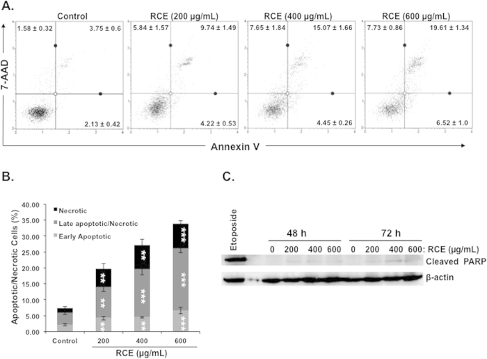
Figure 5. Determination of apoptosis in Rhus coriaria-treated cells.
(A–B) Minimal induction of apoptosis in the MDA-MB-231 cells. Annexin V binding was carried out using Annexin V & Dead Cell kit (Millipore). Cells were treated with or without increasing concentrations of RCE for 48 h. Detached and adherent cells were collected and stained with Annexin V and 7-AAD and then the events for early and late apoptotic cells were counted with the Muse Cell Analyzer as described in Materials and Methods. Data represent the mean ± SEM of at least 3 independent experiments. Statistical analysis was performed using ANOVA followed by LSD Post-Hoc test to determine the significance (*p < 0.05, **p < 0.005, ***p < 0.001). (C) Western blot analysis of PARP cleavage in MDA-MB-231. Cells were treated with increasing concentrations of RCE (200, 400 and 600 μg/mL) for 48 h and 72 h. Exposure of cell to etoposide (50 μM) for 24 h was used as a positive control for apoptosis. The western blots shown are representative of three independent experiments.

