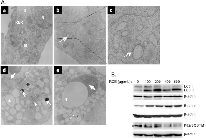
Figure 6. Rhus coriaria induces autophagy in MDA-MB-231 cells.
(A) Representative electron micrographs of untreated MDA-MB-231 cells (a) and MDA-MB-231 cells treated with 400 μM RCE for 48 h (b–e). (B) Western blotting analysis of LC3II, p62(SQSTM1), and Beclin-1 expression RCE-treated MD-MB 231 cells. Cells were treated with or without increasing concentration of RCE for 48 h, then whole cell proteins were extracted and subjected to Western blot analysis, as described in materials and methods, for LC3II, 62(SQSTM1), Beclin1 and β-actin (loading control) proteins. The western blots shown are representative of at least three independent experiments.

