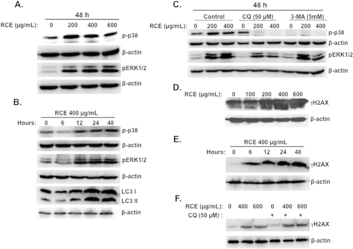
Figure 8. Activation of p38 and ERK1/2 and accumulation of DNA damage in RCE-treated MDA-MB-231 cells.
(A) Concentration-dependent accumulation of phospho-p38 and pERK1/2in RCE-treated cells. MDA-MB-231 cells were treated with and without increasing concentrations of RCE for 48 h and p-p38 and ERK1/2 levels were determined by western blot. (B) Time-course accumulation of p-p38 and pERK1/2 in treated MDA-MB-231 cells. Cells were treated with 400 μg/mL RCE and protein levels of p-p38 and pERK1/2 was determined by western blot at different time-point (6, 12, 24 and 48 h) post-treatment. (C) Effects of autophagy inhibitors on the activation of p-p38 and pERK1/2. Cells were pretreated with or without 3-MA (5 mM) or CQ (50 μM) for 1 h and then RCE was added at the indicated concentrations for 48 h. Proteins were extracted and levels of p-p38 and pERK1/2 was determined by western blot. (D) Concentration-dependent accumulation of γH2AX, marker of DNA damage, in RCE-treated cells. MDA-MB-231 cells were treated with and without increasing concentrations of RCE for 48 h and DNA damage was analyzed, by western blot, by determining the level of γH2AX accumulation using anti- phospho-H2AX (ser 139) antibody. (E–F) DNA damage precedes autophagy. (E) Time-course measurement of DNA damage and LC3-II accumulation in treated MDA-MB-231 cells. Cells were treated with 400 μg/mL RCE and DNA damage and autophagy induction was examined, as described above, at different time-point (6, 12, 24 and 48 h). (F) Effect of autophagy inhibitors on the accumulation of DNA damage. Cells were pretreated with CQ (50 μM) or 3-MA (5 mM) for 1 h before adding RCE (400 and 600 μg/mL) for 48 h. Cells were then harvested and γH2AX level was determined by western blot as described above. The western blots shown are representative of at least three independent experiments.

