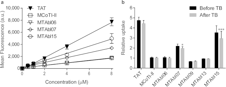
Figure 6. Internalization of MCoTI-II, MTAbl06, MTAbl07, MTAbl09, MTAbl13, and MTAbl15 into HeLa cells.

(a) Peptides were conjugated with one Alexa Fluor® 488 molecule. The mean fluorescence intensity (a.u.) of HeLa cells treated with peptides at varying concentrations for 1 h at 37 °C was analyzed using a flow cytometer with excitation at 488 nm and emission at 530 nm (with 30 nm band pass). The cell-penetrating peptide TAT was included as a positive control. The values plotted in this figure were obtained after the addition of the aqueous soluble quencher trypan blue (TB), which quenches the fluorescence of non-internalized peptides and of cells that have their membranes compromised. (b) The relative cellular uptake to MCoTI-II of cells treated with TAT or five grafted peptides before and after the addition of TB. Internalization of MTAbl peptides into HeLa cells was compared to that of MCoTI-II by comparison of mean fluorescence emission intensity after the addition of TB using one-way ANOVA (*p < 0.05; ***p < 0.001). Results shown here are the mean ± SEM from three independent experiments.
