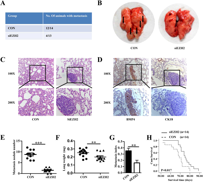
Figure 5. Inhibition of in vivo lung metastasis by EZH2 knockdown.
(A) Number of mice bearing lung metastases. (B) Macroscopic appearances of lung metastasis are shown. Metastatic tumor nodules were identified as whitish and patchy areas. (C) Representative hematoxylin-eosin stained images of the lungs shown in B. (D) Representative IHC staining images of the lungs as shown in B. (E) Number of tumor nodules was evaluated and analysed between MTF cells transfected with lenti-shEZH2 and control group. ***P < 0.001. (F) Lung metastasis index (ratio of tumor area to the total tumor and lung area) was analysed between siEZH2 and the control group. **P < 0.01. (G) Mean weight of the lungs. **P < 0.01. All values in E-G are mean + SD of the 14 mice in siEZH2 CON group and 13 mice in siEZH2 group. (H) EZH2 silencing led to later death of mice seen by survival curve analysis. *P < 0.05, n = 14.

