
Fig. 1.
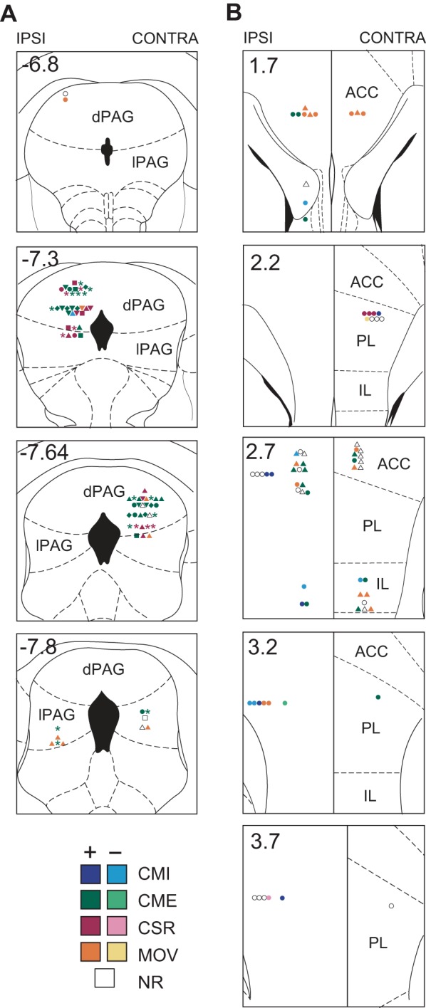
Histological reconstruction of recording sites in dorsolateral periaqueductal gray (dlPAG) and medial prefrontal cortex (mPFC). Reconstructed recording sites of dlPAG neurons (A; n = 74) and mPFC neurons (B; n = 71) are overlaid on coronal templates (with coordinates in mm relative to bregma) from the atlas of Paxinos and Watson (1997). Symbol colors indicate the type classification of each cell (see results), and symbol shapes indicate how the cell responded to conditioned stimulus (CS) pips or shock pulses (*, excited by pip and shocks; ▲, excited by shocks but not pips; ■, excited by pips but not shocks; ⧫, excited by pips and inhibited by shocks, ▼, no pip response and inhibited by shocks; ●, no response to pips or shocks). Left and right sides of the midline correspond to hemispheres ipsilateral (IPSI) and contralateral (CONTRA) to the eyelid where periorbital shocks were delivered. dPAG, dorsal PAG; lPAG, lateral PAG; ACC, anterior cingulate; PL, prelimbic; IL, infralimbic; CMI, conditioned movement inhibition; CME, conditioned movement excitation; CSR, CS responsive; MOV, movement cell; NR, nonresponsive.