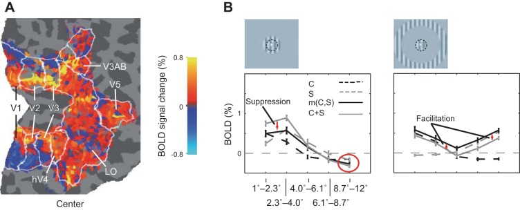
Fig. 3.

Spread of BOLD signal in the primary visual cortex (V1). A: the BOLD signal change without threshold calculated for a central ring (B, left). When no threshold is applied, it is apparent that the BOLD signal spreads first positive and then negative to cover the full extent of the low-level retinotopic areas V1, V2, and V3. B: stimuli consisted of rings containing gratings. The center stimulus is the innermost ring (denoted C, delineated with a black dashed line in the inset), whereas the near surround (S, on the left) and the far surround (right) are the more eccentric ones. Note the consistently negative BOLD at eccentricities several degrees off the primary representation for both C and S stimuli (red circle), the suppression [m(C,S) < C + S] with near surround (red arrows, left) and the trend for facilitation [m(C,S) > C + S] in the far surround condition (red arrows, right) at all eccentricities. C stands for center; S, for surround; m(C,S), for center and surround together; and C + S, for the summation of the BOLD signals measured separately for the C and S conditions. Adapted from Sharifian et al. (2013).
