
Figure 1.
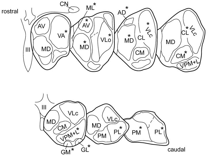
Diagrammatic illustration showing the seven frontal slices, along the rostro-caudal axis, into which each of the control and PD thalami was divided. Delineated and labeled are those 14 nuclear regions from which tissue samples were dissected for noradrenaline analyses. The individual thalamic nuclei taken from a given slice are marked by an asterisk (*). Abbreviations: AD = anterodorsal; AV = anteroventral; CL = centrolateral; CM = centromedian; GL = lateral geniculate; GM = medial geniculate; MD = mediodorsal; ML midline (undivided); PL = lateral pulvinar; PM = medial pulvinar; VA = ventroanterior; VLo = ventrolateral oral; VLc = ventrolateral caudal; VPM+L = ventroposterior medial+lateral (combined). CN = caudate nucleus; III = third ventricle.